
Ficheiro:10.1371 journal.pbio.0030137.g001-L-A.jpg
Dimensões desta antevisão: 800 × 582 píxeis. Outras resoluções: 320 × 233 píxeis | 640 × 465 píxeis | 1 024 × 745 píxeis | 1 280 × 931 píxeis | 2 020 × 1 469 píxeis.
Imagem numa resolução maior (2 020 × 1 469 píxeis, tamanho: 514 kB, tipo MIME: image/jpeg)
|
|
Esta imagem provém do Wikimedia Commons, um acervo de conteúdo livre da Wikimedia Foundation que pode ser utilizado por outros projetos.
|

|
File:Anatomy of Human Ear with Cochlear Frequency Mapping.svg é uma versão vetorial deste ficheiro. Ela deve ser usada em vez desta imagem em formato raster, se não for de qualidade inferior.
File:10.1371 journal.pbio.0030137.g001-L-A.jpg → File:Anatomy of Human Ear with Cochlear Frequency Mapping.svg
Para mais informações, consulte Ajuda:SVG. |
 |
| Descrição |
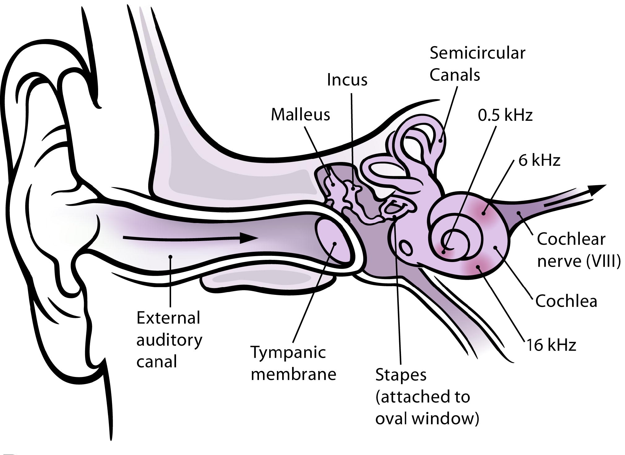
English: The human ear and frequency mapping in the cochlea. The three ossicles incus, malleus, and stapes transmit airborne vibration from the tympanic membrane to the oval window at the base of the cochlea. Because of the mechanical properties of the basilar membrane within the snail-shaped cochlea, high frequencies will produce a vibration peak near the oval window, whereas low frequencies will stimulate receptors near the apex of the cochlea (locations for three frequencies indicated schematically). Information from the cochlear receptor cells is transmitted to the cochlear nuclei via the 8th cranial nerve, and on through the midbrain to the cortex. (Redrawn from Figure 12.3 in [11].) |
| Data |
|
| Origem | |
| Autor |
|
| Esta é uma imagem retocada, o que significa que a versão original foi alterada digitalmente. Modificações: Isolated subfigure A. O original pode ser visto aqui: 10.1371 journal.pbio.0030137.g001-L.jpg:
|
Eu, titular dos direitos de autor desta obra, publico-a com as seguintes licenças:
A utilização deste ficheiro é regulada nos termos da licença Creative Commons - Atribuição 2.5 Genérica.
- Pode:
- partilhar – copiar, distribuir e transmitir a obra
- recombinar – criar obras derivadas
- De acordo com as seguintes condições:
- atribuição – Tem de fazer a devida atribuição da autoria, fornecer uma hiperligação para a licença e indicar se foram feitas alterações. Pode fazê-lo de qualquer forma razoável, mas não de forma a sugerir que o licenciador o apoia ou subscreve o seu uso da obra.
Pode escolher a licença que quiser.
Registo de carregamento original
This image is a derivative work of the following images:
- File:10.1371_journal.pbio.0030137.g001-L.jpg licensed with Cc-by-2.5, Cc-by-2.5
- 2009-02-12T04:06:25Z Mike.lifeguard 2020x2480 (483539 Bytes) {{Information |Description={{en|1=(A) The human ear and frequency mapping in the cochlea. The three ossicles incus, malleus, and stapes transmit airborne vibration from the tympanic membrane to the oval window at the base of
Carregada com derivativeFX
Histórico do ficheiro
Clique uma data e hora para ver o ficheiro tal como ele se encontrava nessa altura.
| Data e hora | Miniatura | Dimensões | Utilizador | Comentário | |
|---|---|---|---|---|---|
| atual | 21h57min de 28 de abril de 2009 | | 2 020 × 1 469 (514 kB) | Mike.lifeguard | malleus and incus were swapped |
| 04h11min de 12 de fevereiro de 2009 |  | 2 020 × 1 469 (521 kB) | Mike.lifeguard | {{Information |Description={{en|1=The human ear and frequency mapping in the cochlea. The three ossicles incus, malleus, and stapes transmit airborne vibration from the tympanic membrane to the oval window at the base of the cochlea. Because of the mechan |
Utilização local do ficheiro
Não há nenhuma página que use este ficheiro.
Utilização global do ficheiro
As seguintes wikis usam este ficheiro:
- eu.wikipedia.org
- sr.wikipedia.org
- zh-yue.wikipedia.org